Highlight
• Alzheimer's disease is the most common neurodegenerative disorder.
• Network analysis can help to identify functional modules and critical genes.
• EOAD risk genes have a role in underlying diseases.
• Control of underlying diseases related to EOAD can prevent AD.
Introduction
Alzheimer's Disease (AD) is the most common neurodegenerative disorder and a complex brain disorder whose risk genes have not been fully identified [1]. Dr. Alois Alzheimer, a German psychiatrist, first identified the disease in 1907 [2]. As one of the main causes of dementia AD, is a major concern for global health, and its consequences have a great impact on individuals and societies [3]. According to the World Health Organization (WHO), AD and other forms of dementia are among the top 10 causes of death in the world, with 65% of deaths from this disease being women. 24 million people worldwide suffer from dementia, with AD constituting the predominant form of this neurodegenerative condition [4]. With the rising average age of the population, it is predicted that the number of people with AD will reach over 81 million by 2040 [5]. The incidence of this disease exhibits exponential growth, and the risk of AD doubles every five years after the age of 65. The main risk factors for AD include genetics, aging, and neuroinflammation. The disease typically begins with memory loss and progresses to cognitive impairment, such as cognitive decline, disorientation, difficulty in independent functioning, and speech impairment, which can interfere with daily activities [6]. Research has shown that low birth weight, preterm birth, and diabetes are related to dementia and AD. Females show a disproportionately higher prevalence of AD for their age, suggesting the influence of additional risk factors beyond lifespan [7]. AD is categorized into two groups depending on the age at which it begins: Early-onset Alzheimer's Disease (EOAD), which occurs before the 65 age, and Late-Onset Alzheimer's Disease (LOAD), which occurs after the 65 age. AD is related to Neurofibrillary Tangles (NFTs) and senile plaques. Amyloid-beta, a major component of senile plaques, has various pathological effects on organelle function and cells. Mutations in three genes PSEN1, PSEN2, and APP are more commonly associated with EOAD by encoding the amyloid beta precursor protein APP and production of amyloid beta [8]. These genes are involved in the amyloid genic pathway, which leads to the formation of amyloid plaques, a hallmark of AD. In LOAD, factors such as lifestyle, environment, genetics, and old age play a significant role, with about 10% of all patients in this group. The APOE gene is the strongest genetic risk factor for LOAD. Other genes, such as BIN1, CD33, and PICALM, have also been associated with an increased risk of developing LOAD. Previous research suggests that as people age, they become more susceptible to neurodegenerative disorders, in which a series of biological events, including severe inflammation, oxidative damage, compromised metabolism, endocrine disruption, and organ decline manifest [9,10]. As AD progresses, individuals experience a relentless erosion of cognitive function that manifests as debilitating memory deficits, delusions, and confusion. Several pathophysiological mechanisms, including the contraction of blood vessels and muscles, dysfunction of mitochondria, and production of free radicals, are involved in memory loss.
APP is a membrane protein that penetrates the membrane of nerve cells and is essential for the growth, survival, and repair of nerve cells. The gene encoding the APP protein is located on chromosome 21. The PS gene is responsible for the production of an intramembrane protein, and mutation in this gene causes a change in the amyloid precursor protein, leading to increased amyloid production. Mutations in both the APP and PS proteins consistently elevate levels of amyloid-beta peptides, particularly the more aggregation-prone amyloid-beta42. Amyloid-beta forms Ca2+ permeable pores and modulates several synaptic proteins, including mGluR5, VGCC, and NMDAR, inducing the overloading of neurons with calcium ions. As a result, cellular Ca2+ disturbances lead to autophagy defects, neuronal apoptosis, mitochondrial abnormalities, synaptic vulnerability, defective neurotransmission, and neurodegeneration in AD. Increased oxidative stress and metabolic disorders in AD lead to Endoplasmic Reticulum (ER) stress and neuronal death. ER stress is a condition caused by an imbalance between protein production and development, leading to problems in protein reactions. FAD-linked PS1 mutation reduces the unfolded protein response and results in vulnerability to ER stress. ApoE protein plays a role in the metabolism of body fats. This protein is involved in AD and cardiovascular disease and carries cholesterol into and out of the cells. Maintaining an optimal cholesterol level in the blood and the balancing role that lipoproteins play in cholesterol transport in the nervous system are important for brain health and neuronal function. In addition to the above genes, using the new GWAS approach (Genome-Wide Association Study), several chromosomal regions and some genetic loci related to the risk of AD have been identified. These regions are responsible for disease signaling and are involved in major pathophysiological pathways such as amyloid precursor protein metabolism. In genomic studies, it was found that 19 gene regions, CASS4, CELF1, FERMT2, HLA-DRB5, INPP5D, MEF2C, NME8, PTK2B, SORL1, ZCWPW1, SLC24A4, CLU, PICALM, CR1, BIN1, MS4A, ABCA7, EPHA1 and CD2AP, are effective in the risk of AD.
Multiple hypotheses have been proposed for the cause of AD. The amyloid beta cascade hypothesis states that amyloid accumulation initiates AD, and evidence shows that disruption in Aβ clearance rather than overproduction of it causes AD. This molecule is the main factor in AD, and the results of mutations in the APP, PSEN1, and APOE genes also support this hypothesis. The amyloid oligomer hypothesis is the other hypothesis, leading to significant progress has been made in understanding the mechanisms of synapse destruction and neuronal death associated with it. Also, due to the role of lysosome destruction and autophagy in AD, other hypotheses for synapse destruction and neuronal death were proposed. Although amyloid-beta is at the forefront of the causative cascade of AD, it is also involved in the formation of hyperphosphorylated tau and NFT formation. Today, it is believed that amyloid beta and tau have mutual relationships and mutual effects. Many molecular pathways that lead to synapse destruction and neuronal death interact with each other, so a single hypothesis for the molecular causes of AD cannot be proposed.
AD is related to several diseases, such as type 2 diabetes, Down syndrome, bowel diseases, and kidney diseases. Research shows that diabetes and any other disease that affects blood sugar can affect brain and nervous system function. Increased blood sugar levels increase amyloid-beta levels in the patient's body, which can lead to AD. Down syndrome is one of the most common genetic disorders that is caused by the complete or partial trisomy of chromosome 21. In this trisomy, people with Down syndrome have an extra copy of the APP gene and produce too much amyloid-beta. Almost all adults with Down syndrome develop amyloid plaques and neurofibrillary tangles, which are signs of AD. Furthermore, with improvements in healthcare and longer lifespans for individuals with Down syndrome, the population diagnosed with AD within this community is steadily increasing. These results show the need for further research and early intervention strategies to address the specific challenges of AD in people with Down syndrome. Recently, the relationship between constipation and changes in the gut microbiome with AD has been identified. Changes in the gut microbiome have been reported as inflammatory responses in the development of AD. This inflammatory response leads to damage to the central nervous system. In addition, metabolites and cytokines are secreted from the intestinal cells, which affect the activity of the microbiome and affect the activity of neurons. Recent studies have identified an association between Chronic Kidney Disease (CKD), particularly in elderly populations, and an increased risk of developing various forms of cognitive decline, ranging from Mild Cognitive Impairment (MCI) to dementia.
These findings have helped to identify better the biological pathways involved in the disease, and the promising results from this study suggest future therapies might be developed to treat or prevent AD. Although there is no known way to stop or prevent the progression of this disease, some treatments help to improve the symptoms of the disease. Given the increasing number of people affected, it is very important to identify the metabolic pathways involved in the disease and predict target genes and biomarkers to help early diagnosis of the disease. In this study, we try to use bioinformatics tools to investigate the genes identified for AD and analyze gene interactions, and metabolic pathways.
Materials and Methods
Data collection
First, genes related to AD were identified and selected by searching in the NCBI, and DisGeNET database.
Construction and analysis of protein-protein interaction network
The identified genes were uploaded to the STRING database (version 11.5) and the Protein-Protein Interaction (PPI) network of AD genes was constructed. Then, to analyze the topology of the AD network, key genes, and functional modules were identified using the Cytoscape software (version 3.9.1). The MCODE plug-in (Molecular complex detection) with default settings (K-Core:2, Node Score Cutoff:0.2, Max Depth:100 Haircut: True, and DegreeCutoff:2) was used for clustering the network and identifying functional modules. Cytohubba plug-in based on the MCC ranking method was used to identify key genes in the AD PPI network.
Gene network enrichment
To identify GO terms include Biological Process (BP), Cellular Component (CC), and Molecular Function (MF) using g: Profiler. In addition, an investigation of the metabolic pathways of AD was used in the Kyoto Encyclopedia of Genes and Genomes (KEGG). The microRNAs (miRNAs) regulate many cellular processes and metabolic pathways and have a role in various diseases, including neurological diseases, cancer, and inflammatory diseases. So, in this study to identified miRNAs associated with key genes and AD, using the Mirwalk and MirNet web servers, respectively. The workflow of this study is shown in Figure 1.
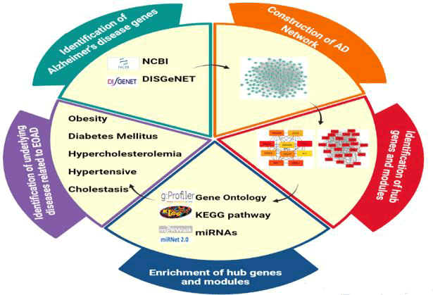
Figure 1. Research workflow.
Results
Construction and analysis of protein network
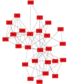
A number of 200 genes were identified for AD using NCBI and DisGeNET database. Then AD genes were submitted to the STRING database and constructed the AD network. This network contains 1105 edges, average node degree=13.3, and PPI enrichment P-value<1.0e-16, which is shown in Figure 2.
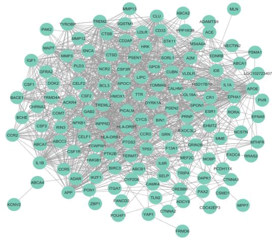
Figure 2. AD gene network.
GO enrichment analysis of modules
The AD PPI network was clustered using the MCODE plug-in, and related genes were placed in five functional modules. The functional enrichment analysis of BP for each module was investigated using the g: Profiler. The results are presented in Table 1.
| Module name |
Node number |
Node name |
GO ID |
GO function |
Image of the module |
| M1 |
23 |
FERMT2, PSEN1, SORL1, EPHA1, PLD3, APOE, PSEN2, CD2AP, RIN3, PTK2B, ZCWPW1, PICALM, CELF1, BIN1, MEF2C, HLA-DRB5, MS4A6A, ABCA7, CD33, CLU, INPP5D, HLA-DRB1, TREM2 |
GO:0034205
GO:0051239 GO:0070374 GO:0043523 GO:0007611 |
Amyloid-beta formation,
Regulation of multicellular organismal process,
Positive regulation of ERK1 and ERK2 cascade,
Regulation of neuron apoptotic process,
Learning or memory |
 |
| M2 |
16 |
TP53, SQSTM1, PTGS2, CSF3, BCHE, A2M, IL6R, CSF2, CSF1, IL1A, CYCS, NFKB1, GRN, MME, PRNP, IGF1 |
GO:0051247 GO:0010033 GO:0050790 GO:0010638 GO:0007265 |
Positive regulation of protein metabolic process,
Response to organic substance,
Regulation of catalytic activity,
Positive regulation of organelle organization,
Ras protein signal transduction |
 |
| M3 |
26 |
SNCA, LIPC, GRIN2B, PON1, TTR, BCL3, HMGB1, NCSTN, CTSD, LDLR, CSF3R, ABCA1, MMP3, APP, CCR2, IDE, CSF1R, MMP12, IL1B, CCR3, SELP, APOC1, TYROBP, VLDLR, MMP13, BIRC3 |
GO:0010033 GO:0008203
GO:0097006 GO:0006897
GO:0030301 |
Response to organic substance,
Cholesterol metabolic process,
Regulation of plasma lipoprotein particle levels,
Endocytosis,
Cholesterol transport |
 |
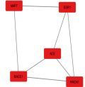
| M4 |
5 |
HMOX1, ESR1, ACE, MAPT, BAC1 |
GO:1901700 GO:0010288 GO:0140448
GO:1990000
GO:0009628 |
Response to oxygen-containing compound,
Response to lead ion,
signaling receptor ligand Precursor processing,
Amyloid fibril formation,
Response to abiotic stimulus |
 |
| M5 |
3 |
MTHFR, ABCB1, COMT |
GO:0009410 |
Response to xenobiotic stimulus |
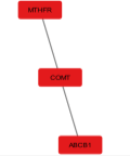 |
Table 1. GO enrichment of functional modules M1 to M5.
The metabolic pathways of AD in the KEGG will be shown in Figure 3.
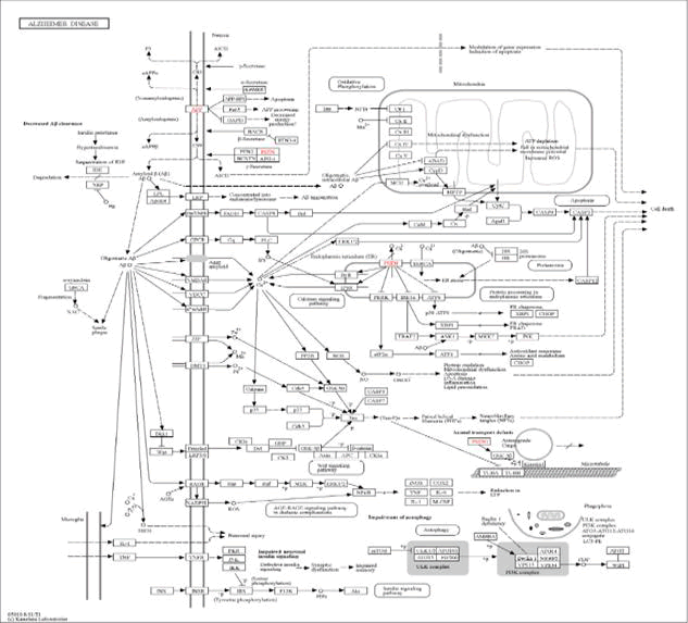
Figure 3. Metabolic pathways of AD in KEGG-map05010. In the metabolic pathways of AD, APP, APOE, PS1, and PS2 genes, which are shown in red, play a role in the formation of amyloid plaques, oxidative stress, inflammation, calcium disturbances, and other metabolic disorders.
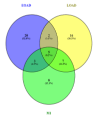
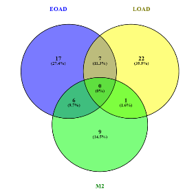
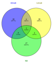
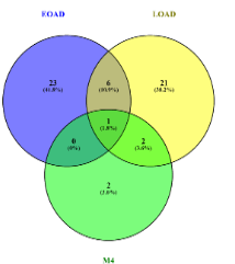
Also, after the investigation of each module, the common genes between them and EOAD and LOAD were found and reported in Table 2.
| Module name |
EOAD genes |
LOAD genes |
Common EOAD and LOAD genes |
Fig |
| M1 |
PSEN2, PLD3, RIN3 |
BIN1, CLU, CD33, PICALM, EPHA1, CD2AP, PTK2B |
PSEN1, APOE, SORL1, TREM2, ABCA7 |
 |
| M2 |
CSF1, CSF3, IL1A, CSF2, PRNP, NFKB1 |
A2M |
- |
 |
| M3 |
SNCA, TYROBP, CSF1R, CSF3R, NCSTN |
IDE, LDLR, ABCA1 |
APP |
 |
| M4 |
- |
ACE, BACE1 |
MAPT |
 |
| M5 |
- |
MTHFR |
- |
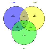 |
Table 2. The common genes between each module and EOAD and LOAD.
Key genes analysis
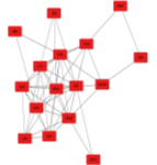
Ten key genes were identified in the AD gene network. These key genes are containing APOE, TREM2, SORL1, BIN1, PICALM, ABCA7, CD2AP, CD33, MS4A6A, and CLU. The interaction network of 10 key genes is shown in Figure 4.
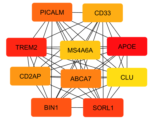
Figure 4. AD key genes Network.
GO enrichment analysis of key genes
Functional enrichment of key genes was investigated using the g: Profiler platform, and the results are presented in Table 3.
| GO ID |
GO terms |
Function |
| GO:0001540 |
MF |
Amyloid-beta binding |
| GO:0048156 |
MF |
Tau protein binding |
| GO:0050750 |
MF |
Low-density lipoprotein particle receptor binding |
| GO:0071813 |
MF |
Lipoprotein particle binding |
| GO:0019828 |
MF |
Aspartic-type endopeptidase inhibitor activity |
| GO:0019899 |
MF |
Enzyme binding |
| GO:0005543 |
MF |
Phospholipid binding |
| GO:1902992 |
BP |
Negative regulation of amyloid precursor protein catabolic process |
| GO:0097242 |
BP |
Amyloid-beta clearance |
| GO:0051050 |
BP |
Positive regulation of transport |
| GO:0061024 |
BP |
Membrane organization |
| GO:1902532 |
BP |
Negative regulation of intracellular signal transduction |
| GO:0051241 |
BP |
Negative regulation of multicellular organismal process |
| GO:0097006 |
BP |
Regulation of plasma lipoprotein particle levels |
| GO:0007611 |
BP |
Learning or memory |
| GO:0061518 |
BP |
Microglial cell proliferation |
| GO:0019216 |
BP |
Regulation of lipid metabolic process |
| GO:0036477 |
CC |
Somatodendritic compartment |
| GO:0032994 |
CC |
Protein-lipid complex |
| GO:0009986 |
CC |
Cell surface |
| GO:0005794 |
CC |
Golgi apparatus |
| GO:0097418 |
CC |
Neurofibrillary tangle |
| GO:0071944 |
CC |
Cell periphery |
| GO:0030054 |
CC |
Cell junction |
| GO:0005771 |
CC |
Multivesicular body multivesicular body |
| GO:0099020 |
CC |
Perinuclear endoplasmic reticulum lumen |
Table 3. Top 10 GO terms of key genes.
Identification of underlying diseases
Investigation of key genes in DisGeNET showed that five key genes that contain BIN1, CLU, CD33, PICALM, and CD2AP are related to LOAD, and four key genes that contain APOE, SORL1, TREM2, and ABCA7 are playing a role in EOAD. TREM2 and ABCA7 genes play a critical role in the development of neurodegenerative diseases such as Alzheimer's disease, Parkinson's, memory impairment, dementia, and cognitive impairment. The APOE and SORL1 genes, have a direct role in AD, and also, play an indirect role in the occurrence of this disease. APOE is one of the genes that cause obesity, hypercholesterolemia, diabetes mellitus, and hypertensive disease through the metabolism, signal transduction, transport of small molecules, metabolism of proteins, vesicle-mediated transport, gene expression (transcription), and sensory perception pathways. Also, SORL1 genes cause obesity, cholestasis, diabetes mellitus (non-insulin-dependent), and hypercholesterolemia through the metabolism of protein pathways.
miRNA-key gene interaction network
MicroRNAs (miRNAs) are short RNA molecules that consist of 23-17 nucleotides. They play a key role in regulating gene function. These molecules interact with target molecules that have a critical role in protein synthesis. In this study, the miRNAs associated with key genes and AD were extracted using the miRWalk and MirNet, respectively. 156 miRNAs were found to be shared between the AD and key genes. The miRNA-key gene interaction network was constructed using Cytoscape (Figure 5). This result shows the importance of the key genes in AD disease.
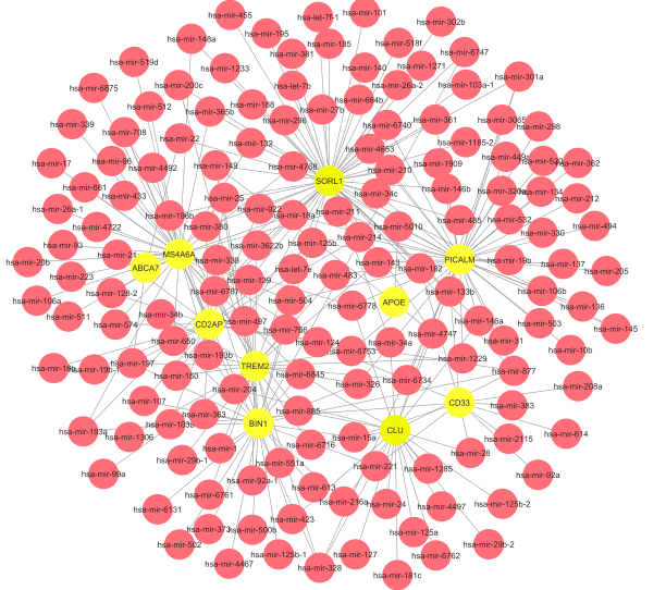
Figure 5. miRNA-key gene interaction network in AD.
Discussion
After analyzing the AD gene network, the functional module and key genes were identified and then the functional enrichment analysis of them was performed. Five functional modules were identified, and each of them has an important role in different processes of AD. The function of each module is as follows:
Module M1 consists of 23 nodes and all of the 10 key genes were found to be located in this Module. M1 has a critical role in the amyloid-beta formation, regulation of multicellular organismal process, positive regulation of ERK1 and ERK2 cascade, regulation of neuron apoptotic process, and learning or memory. In this module, three genes contain PSEN2, PLD3, and RIN3 except in EOAD and seven genes contain BIN1, CLU, CD33, PICALM, EPHA1, CD2AP, and PTK2B except in LOAD. Also, five genes including PSEN1, APOE, SORL1, TREM2, and ABCA7 are shared between EOAD and LOAD. This result shows the importance of this module in AD.
Module M2 with 16 genes. M2 is involved in positive regulation of protein metabolic process, response to organic substance, regulation of catalytic activity, positive regulation of organelle organization, and Ras protein signal transduction. Six genes containing CSF1, CSF3, IL1A, CSF2, PRNP, and NFKB1 are EOAD genes and one gene that contains A2M is in the LOAD list of genes. These results show that M2 is mostly involved in EOAD.
Module M3 contains 26 genes. M3 has an important role in response to organic substances, cholesterol metabolic process, regulation of plasma lipoprotein particle levels, endocytosis, and cholesterol transport. In this module, five genes containing SNCA, TYROBP, CSF1R, CSF3R, and NCSTN are EOAD genes and three genes containing IDE, LDLR, and ABCA1 are LOAD genes. Also, the APP gene is common between EOAD and LOAD. It seems this module is related to EOAD.
Module M4 contains five genes. This module is related to in response to oxygen-containing compounds, response to lead ions, signaling receptor ligand precursor processing, amyloid fibril formation, and response to abiotic stimulus. Two genes that contain ACE, and BACE1 in LOAD and MAPT genes are common between EOAD and LOAD.
Module M5 has 3 genes. M5 is involved in the response to xenobiotic stimulus. The MTHFR gene in this module is one of the LOAD genes.
Considering these results, it seems to be that M2 and M3 are related to EOAD and M4, and M5 is related to LOAD.
Identifying key genes and their vital pathways can help us to improve our understanding of diseases. In this study, we identified 10 key and the results are as follows:
APOE gene: APOE protein is one of the most important apolipoproteins in the brain and an amyloid-associated protein of AD. It is involved in the transport of cholesterol and other lipids. The ApoE gene exists in three forms that include ApoE2, ApoE3, and ApoE4. The ApoE4 gene leads to increased development of AD risk and is known to be an important factor in the formation and accumulation of amyloid plaques in the brain.
BIN1 gene: This gene encodes a protein that is essential for the formation of cellular structures. BIN1 gene variants in combination with APOE are associated with an increased AD risk. The BIN1 gene has a role in endocytosis and intracellular trafficking like in the neurofibrillary tangles and formation of β amyloid plaques, which are important factors in the pathological of AD.
PICALM gene: PICALM is an amyloid-associated protein that is involved in cellular endocytosis processes. Mutation in this gene is associated with an increased risk of developing AD. GWAS studies have identified the PICALM gene as the most important source of genetic susceptibility after APOE and BIN1. This gene encodes a protein essential for endocytosis, and its effect as an AD risk gene is confirmed across variant populations through independent genetic studies. The progression of tau pathology has also been confirmed in AD models. Interestingly, the predominant expression of PICALM in the brain's small blood vessels suggests a unique and potentially crucial role in cerebrovascular function and AD pathogenesis.
ABCA7 gene: This gene is an amyloid-associated protein that is responsible for the transport of phospholipids in cells. It has a significant role in early-onset dementia and amyloid deposition. Also, recent GWAS studies show that the ABCA7 variants are risk factors for LOAD.
CD2AP gene: CD2AP is an amyloid-associated protein that is involved in calcium production, synaptic structure, and function of the brain. Cell-to-cell interactions (specific phospholipids) and amyloid β production, limit the regulation of endocytosis or the processing of amyloid β precursor protein. Evidence has shown that the protein associated with CD2AP is involved in signal transduction and regulation of cellular skeleton molecules. Variations within this gene, known as Single Nucleotide Polymorphisms (SNPs), are related to a higher risk of developing AD.
CD33 gene: It is known as a type I transmembrane protein belonging to the immunoglobulin-like lectins that bind to sialic acid, which mediates cell-cell interactions and inhibits the normal function of immune cells. This gene is mainly expressed in microglial cells. Increased CD33 expression is observed in the brains of Alzheimer's patients, and this increase is positively related to amyloid plaque load and disease severity. In addition, CD33 overexpression disrupts Aβ metabolism and promotes the formation of amyloid plaques in the brain.
SORL1 gene: The SORL1 gene is one of the genes that have a critical role in developing AD. Recent studies show that loss of SORL1, and mutations in APP and PSEN1/2 genes, are associated with AD. The SORLA gene was expressed in neurons of the central nervous system that contain the cortex, cerebellum, hippocampus, and spinal cord. The SORLA mechanism as a neuronal sorting receptor is to control the amyloidogenic processes in the brain. Recent studies show that SORL1 is considered a biomarker in AD.
MS4A6A gene: The MS4A6A gene is in the MS4A family that is involved in the function of brain cells. Its variants with the APOE gene affect the signaling related to AD. Extensive genome-wide studies have identified new loci and functional pathways that are effective in reducing AD risk, which is related to the effects of this gene.
TREM2 gene: This gene can increase the development of AD risk in an individual by up to three times. TREM2 is a transmembrane receptor that is expressed in amyloid cells, and its relation to AD is in the immune and inflammatory pathways. TREM2 plays a critical role in changing the behavior of microglial cells, including their response to amyloid plaques.
CLU gene: Amyloid-associated protein that protects neurons. The protein encoded by this gene is a chaperone that can also be found in the cell under stressful conditions. It is involved in several fundamental biological events such as tumor progression, cell death, and neurological disorders.
GO enrichment analysis showed that the most important biological processes associated with these key genes are the negative regulation of the amyloid precursor protein catabolic process, amyloid-beta clearance, positive regulation of transport, and membrane organization. A significant function of these genes is amyloid-beta binding, tau protein binding, and low-density lipoprotein particle receptor binding, and also, localized mostly in somatodendritic compartment, protein-lipid complex, and cell surface. The miRNA-key gene interaction network showed that half of the identified miRNAs related to AD affect AD by controlling the expression of identified key genes, and this indicates the important role of key genes in the development of AD.
Conclusion
According to the underlying diseases related to EOAD, many studies have been conducted, for example, there are several studies investigating the metabolic and cognitive effects of obesity and the APOE gene but the relationship between APOE and obesity to regulate the pathogenesis of AD is still unclear. Jones N.S. and G.W. Rebeck's study shows the APOE4 gene and obesity increase the risk of developing AD. Also, Zhao T, et al. study shows that APOE is a strong genetic risk factor for AD, and obesity is related to AD. Sparks DL, study shows that hypertension is one of the seasons that cause Senile Plaques (SP) in neurofibrillary tangles and SP is related to Alzheimer's Disease. Also, they show that APOE increases the risk of hypertension, and AD. Xu C, et al. study shows that hypercholesterolemia increases AD pathology by the accumulation of the amyloid-β, and on the other hand, APOE is related to hypercholesterolemia. Patel VN, et al. study shows that AD and type-2 diabetes mellitus are related together and also the diabetes and ApoE increase the risk of AD by neuritic plaques, aggregation neurofibrillary tangles, and amyloid-β.
In recent study shows the association between SORL1 and obesity. Schmidt, V., et al. study shows that SORL1 expression affects obesity and glucose tolerance. This study shows the relationship between SORL1 and obesity leads to neurodegeneration and metabolism that cause AD. Yu Y, et al. study show type 2 diabetes mellitus is related to an increased risk of dementia, like AD. Also, this study shows the SORL1 gene is a critical gene for AD. Yu Y, Feng X, et al. study shows the SORL1 gene is a candidate gene in lipid metabolic pathways in AD. The Nisha V and CS Kumar study shows that SORLA is the underlying cause of AD and is related to metabolic pathologies like obesity, diabetes, and hypercholesterolemia.
The findings of this study showed that bioinformatics tools and systems biology approaches are capable of studying multi-gene diseases such as AD. In addition, key genes, vital pathways, and functional modules can be helpful to the identification of diseases like EOAD to find biomarkers, drug targets, and related diseases. The results of our study showed that obesity, hypercholesterolemia, diabetes mellitus, hypertension, and cholestasis have a critical role in the occurrence or development of EOAD. Therefore, the treatment and control of these diseases can play an important role in preventing EOAD. However, more experimental studies are necessary to confirm the roles of identified key genes, and underlying diseases in the occurrence of AD.
Ethics Approval and Consent to Participate
Not applicable.
Consent for Publication
Not applicable.
Data and/or Code Availability
Not applicable.
Availability of Data and Materials
Data used in this study are available upon request from the corresponding author.
Declaration of generative AI and AI-assisted technologies in the writing process not applicable.
Competing Interests
The authors have no competing interests to declare that are relevant to the content of this article.
Funding
This study received no specific grant from any funding agency in the public, commercial, or not-for-profit sectors.
Authors' Contributions
Kazemi, Zangi Darestani, and Ziaastani made the concept. Zangi darestani and Ziaastani have equal contributions. Zangi darestani and Ziaastani provided the data curation and made the investigation and formal analysis. Kazemi, Abbasnejad, and Kalantari-Khandani supervised the manuscript. Zangi darestani, and Ziaastani wrote the original draft of the manuscript. Kazemi, Abbasnejad, and Kalantari-Khandani reviewed and edited the manuscript. Zangi darestani and Ziaastani prepared the figures. All authors reviewed the manuscript.
Information Sharing Statement
Data sharing does not apply to this article as no new data were created in this study. In this study, we use the NCBI (https://www.ncbi.nlm.nih.gov/), and DisGeNET (https://www.disgenet.com) databases.
Acknowledgments
Not applicable.
References
- Lin CX, Li HD, Deng C, Erhardt S, Wang J, et al. AlzCode: A platform for multiview analysis of genes related to Alzheimer’s disease. Bioinformatics. 2022;38(7):2030-2032.
[Crossref] [Google Scholar] [PubMed]
- Lee JM, Kim SR. Prothrombin kringle-2, a mediator of microglial activation: New insight in Alzheimer’s disease pathogenesis. Neural Regen Res. 2022;17(12):2675-2676.
[Crossref] [Google Scholar] [PubMed]
- Lane CA, Hardy J, Schott JM. Alzheimer's disease. Eur J Neurol. 2018;25(1):59-70.
[Crossref] [Google Scholar] [PubMed]
- Ballard C, Gauthier S, Corbett A, Brayne C, Aarsland D, et al. Alzheimer's disease. Lancet. 2011;377(9770):1019-1031.
[Crossref] [Google Scholar] [PubMed]
- McGirr S, Venegas C, Swaminathan A. Alzheimer’s disease: A brief review. J Exp Neurol. 2020;1(3):89-98.
[Crossref] [Google Scholar]
- Huang LK, Chao SP, Hu CJ. Clinical trials of new drugs for Alzheimer disease. J Biomed Sci. 2020;27:1-3.
[Crossref] [Google Scholar] [PubMed]
- Vina J, Lloret A. Why women have more Alzheimer's disease than men: Gender and mitochondrial toxicity of amyloid-β peptide. J Alzheimer’s Dis. 2010;20(2):527-533.
[Crossref] [Google Scholar] [PubMed]
- Atri A. The Alzheimer’s disease clinical spectrum: Diagnosis and management. Med Clin North Am. 2019;103(2):263-293.
[Crossref] [Google Scholar] [PubMed]
- Li S, Xiao J, Huang C, Sun J. Identification and validation of oxidative stress and immune-related hub genes in Alzheimer’s disease through bioinformatics analysis. Sci Rep. 2023;13(1):657.
[Crossref] [Google Scholar] [PubMed]
- Ju Y, Tam KY. Pathological mechanisms and therapeutic strategies for Alzheimer’s disease. Neural Regen Res. 2022;17(3):543-549.
[Crossref] [Google Scholar] [PubMed]